

Modern Diagnosis of Retinal Diseases
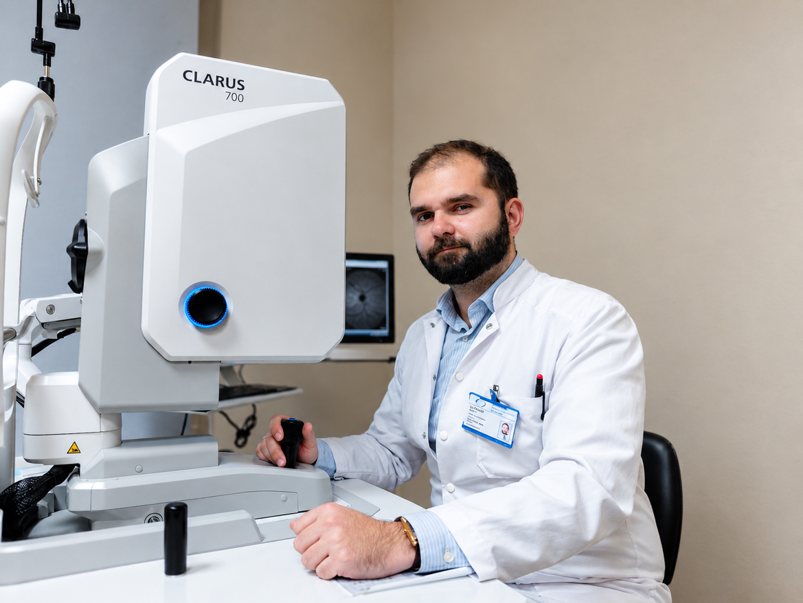
Author: Dr. Milen Lazov
Since ancient times, civilizations have associated the human eye with a variety of beliefs and interpretations. This perspective is entirely consistent with its complex structure. Today, the prevailing view is that of the eye as an optical phenomenon. But it is far more than that. While the cornea and lens focus the image, the retina is the highly specialized sensor that perceives and creates it. The macula, the central but most complex and vulnerable part of the retina, provides central and color vision; the peripheral retina is responsible for light perception and our peripheral vision. Hegel described the world as colorless and formless outside the sense that perceives these qualities.
Types of Retinal Diseases
The retina’s high degree of specialization, combined with its complex structure and extensive capabilities, means that a variety of diseases can affect it. Retinal diseases can be classified as:
- vascular, congenital, and acquired diseases of the macula;
- degenerative conditions of the peripheral retina;
- retinal detachment;
- benign and malignant retinal tumors.
Development of Diagnostic Methods
Since ancient times, the fundus of the eye had been a territory beyond human knowledge. Scientific and technological progress has made it possible to discover and understand it, and even more—to treat its diseases. This entire process ranges from taking a medical history, through early forms of ophthalmoscopy and the advent of fluorescein angiography, to modern diagnostic advances in the era of optical coherence tomography /OCT/.
Together, these provide our strongest weapons in the increasingly successful fight against retinal diseases: prevention, early diagnosis, early treatment, and minimized consequences.
Symptoms and Medical History in Retinal Diseases
The first step in the diagnostic process is taking a focused, detailed, and meaningful medical history. In macular diseases, the patient may primarily report reduced vision, impaired color perception, reduced contrast sensitivity, image distortion /metamorphopsia/, and central visual field defects.
With regard to peripheral involvement, common symptoms include sudden onset of floaters, flashes of light even with the eyes closed (photopsias), and missing areas of the visual field (scotomas). The patient’s family history should not be overlooked, as it is particularly important in hereditary conditions, macular degeneration, and other diseases.
Clinical examination and ophthalmoscopy
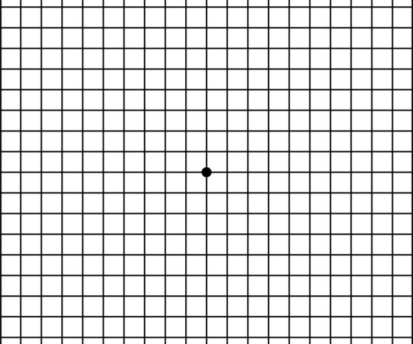
The examination always begins with measurement of best-corrected visual acuity. Diagnoses such as RCS may be suspected at this stage. The Amsler grid is a useful, simple, and readily accessible test that reveals macular damage through distortion of central images. Testing adaptation to light and darkness is useful in some degenerative diseases of the macula and peripheral retina, particularly hereditary ones.
This is followed by an examination of the anterior and posterior segments. This almost always reveals a diagnosis or at least points toward one. Sometimes an examination without dilating the pupils is sufficient, but good practice and a complete examination require pupil dilation. Today, this is achieved with fast-acting, short-duration eye drops. This is critical and important for screening every patient for asymptomatic peripheral retinal degenerations, which, if left untreated, can lead to retinal detachment and irreversible vision damage. The same applies to screening for benign and malignant tumors of the choroid and retina.
Modern tests for retinal diseases
Current tests used for retinal diseases include: color fundus photography, fluorescein angiography, fundus autofluorescence imaging, optical coherence tomography (OCT), better known as an eye scan; ocular ultrasonography;
Color fundus photography
Color fundus photography is useful for documenting findings and monitoring them over time—for example, changes in the size of a tumor.
Fluorescein angiography
Fluorescein angiography is an indispensable gold standard for examining the function of eye structures, particularly the blood-retinal barriers (the pigment epithelium and blood vessel walls). It plays a key role in identifying vascular conditions such as diabetic retinopathy or vascular occlusions; classifying and detecting wet AMD; and evaluating optic nerve disorders, inflammatory diseases of the choroid, and tumors in the fundus. It also allows vascular changes to be staged and areas for laser treatment to be marked.
The contrast dye is virtually harmless and contains no heavy chemical elements. The examination is performed using a specialized high-tech camera, whose “flash” illuminates the dye in the blood vessels at the back of the eye, causing it to fluoresce and emit light that the camera captures as a black-and-white image. Changes are observed over time during the different phases of contrast circulation.
Autofluorescence examination
The principle of autofluorescence imaging is similar, except that it relies on the natural fluorescence of lipofuscin in the tissues. It is particularly useful for staging and monitoring dry AMD, atrophic changes in the macula associated with hydroxychloroquine use, and diagnosing eye tumors.
Optical coherence tomography (OCT)
Perhaps the most significant tool in the modern ophthalmologist’s arsenal is optical coherence tomography (OCT), or an eye scan. The examination is virtually completely safe because the scan uses light rays. OCT provides detailed information about the anatomy of retinal structures.
It is indispensable for various forms of macular degeneration, macular edema of various causes, and vitreoretinal interface disorders, including epiretinal membranes, macular holes, and retinal detachment, where it is crucial for staging and planning the surgical approach and technique; and for diagnosing eye tumors. The vascular module, which works on the principle of the optical Doppler effect, provides information about retinal blood circulation and sometimes offers a noninvasive alternative to fluorescein angiography, but is generally less informative.
It plays a key role in diagnosing wet AMD by visualizing vascular membranes. We use OCT to monitor the progression of all the conditions listed above in detail. OCT is immensely important in diagnosing and monitoring glaucoma and optic nerve disorders.
All the methods listed so far can be used only when the ocular media are clear. When the cornea is cloudy, a cataract is advanced, or the vitreous is opaque, our primary method is ultrasound.
The latest generations of ocular ultrasound devices have sophisticated multicomponent transducers and advanced software, allowing the structures of the eye to be imaged at extremely high resolution. Ultrasound is used for routine examination of eyes with opaque media, retinal and choroidal detachments, diagnosis of eye tumors, eye hemorrhages, and inflammatory eye diseases. It is also used to calculate intraocular lens power in combined surgery (so-called B-biometry).
Why do we combine different examination methods?
Of course, all of these imaging methods have advantages and disadvantages, so they are often used together to complement one another. This allows us to obtain both structural and functional information. For example, in diabetic retinopathy, OCT can monitor macular edema, while fluorescein angiography (FA) can visualize circulatory abnormalities, peripheral edema, abnormal vascular connections, and new blood vessels.
Artificial Intelligence in the Diagnosis of Retinal Diseases
From a provocative novelty to an everyday concern, the use of artificial intelligence is now a practical reality. At present, the main focus is on training AI algorithms to recognize pathological findings. This is being used in screening campaigns in remote areas where medical personnel are scarce. This is a prelude to a second future approach: conducting a screening or standard examination with a human operator or one incorporated into the machine; the examination is sent to a specialist, who analyzes it and determines the next steps.
The availability of so many advanced, informative, and complementary imaging methods undoubtedly makes an accurate, rapid diagnosis that can be tracked over time possible. But the truly significant advantage of ophthalmic diagnostics lies in preventive medicine, actively looking for retinal disease, and responding early when symptoms appear. Early and accurate diagnosis means effective treatment, which is the path to preserving vision. And vision is life.
*This article is part of the awareness campaign “See What Cannot Be Seen,” dedicated to retinal diseases, which we are conducting in partnership with the Retina Bulgaria Association.

Replacing Glasses with Contact Lenses – What is Important to Know
During the summer, many people who wear prescription glasses choose to replace them with contact lenses because this way they.
Read More
Dr. Veselin Daskalov: More than 40,000 cataract surgeries are performed annually in Bulgaria
An ophthalmologist with many years of experience is the guest in this episode of the Bulgarian National Radio podcast “At.
Read More