

Retinal Vascular Diseases
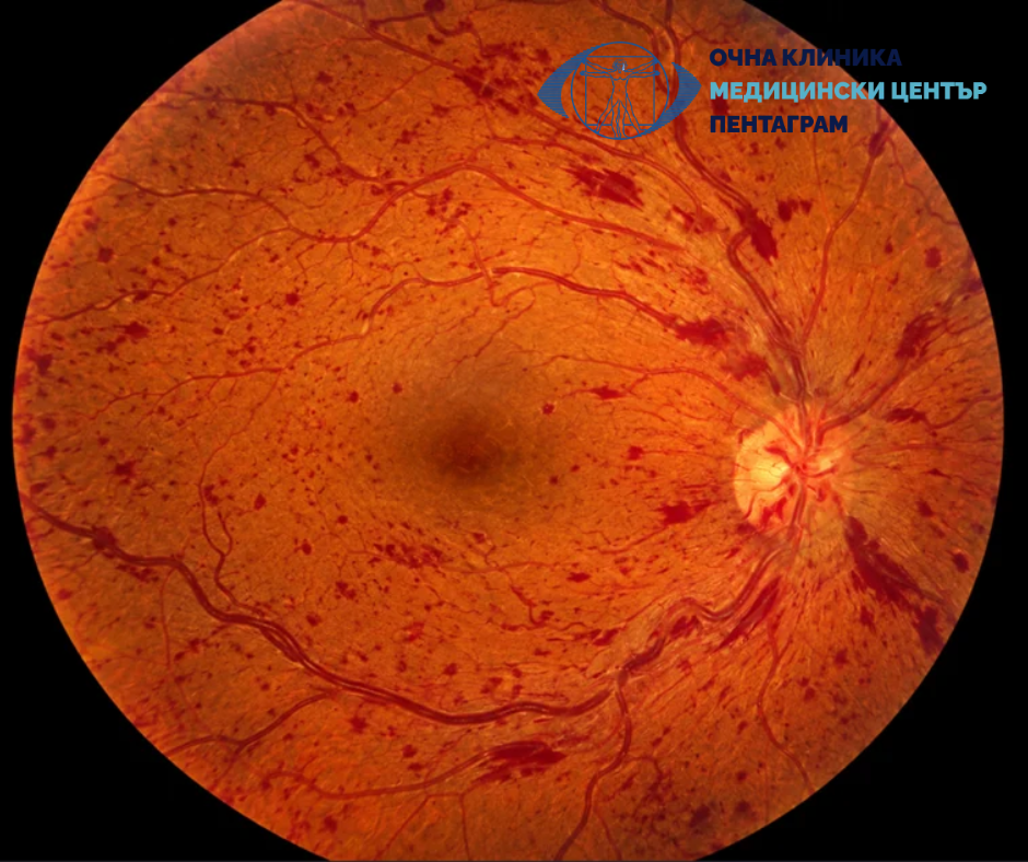
Author: Dr. Doli Efremova

The retina is the innermost layer of the eye. It converts light into nerve signals and allows us to see the world around us. To perform this function, it needs a constant and adequate blood supply. Disruptions to this blood supply can lead to serious vision damage, sometimes without early warning symptoms.
Retinal vascular diseases are associated with some of the most common chronic conditions, including diabetes mellitus and high blood pressure. Some of these diseases, such as blockage (occlusion) of a retinal artery, can occur suddenly and constitute a medical emergency.
Early detection, accurate diagnosis, and timely treatment are essential to preserving vision. Let’s look at the main retinal vascular diseases, how they present, and the available treatment options.
How does the retina receive its blood supply?
The retina is nerve tissue with exceptionally high metabolic demands. To function normally, it requires a continuous supply of oxygen and nutrients and effective removal of waste products.
The blood supply comes from two main sources:
- Retinal circulation – the central retinal artery and its branches primarily supply the inner layers of the retina.
- Choroidal circulation – the extensive network of vessels in the choroid supplies oxygen and nutrients to the outer layers of the retina, including the photoreceptors.
The blood-retinal barrier also plays an important role. It regulates the passage of substances from the blood into retinal tissue and maintains a stable environment for nerve cells to function.
When blood flow is disrupted or the vascular barrier becomes permeable, ischemia, hemorrhages, and fluid accumulation and swelling can occur. If the macula is affected, visual function can deteriorate significantly.
What are the main retinal vascular diseases?
Diabetic retinopathy
Diabetic retinopathy is one of the leading causes of blindness worldwide. It develops as a result of prolonged damage to the small blood vessels of the retina caused by high blood sugar levels. In the early stages, the disease may cause no symptoms despite the presence of changes in the retina.
There are two main forms:
- Nonproliferative diabetic retinopathy – characterized by microaneurysms, intraretinal hemorrhages, hard exudates, and varying degrees of ischemic changes.
- Proliferative diabetic retinopathy – significant ischemia stimulates the formation of new, abnormal blood vessels, known as fibrovascular proliferation. This can cause bleeding into the vitreous and tractional retinal detachment.
A particularly important complication is diabetic macular edema. In this condition, fluid accumulates in the macula, the area responsible for central vision and seeing detail. Patients may notice blurring, distortion of images, or difficulty reading.
The risk of developing diabetic retinopathy and of its progression increases with the duration of diabetes and with poor blood sugar control, high blood pressure, and lipid disorders.
What is important to know? The absence of vision problems does not mean that the retina is healthy. People with diabetes should have regular eye exams with pupil dilation, usually at least once a year, and more often if changes are detected, as recommended by their ophthalmologist.
Retinal vein occlusions
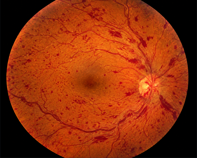
Retinal vein occlusion is a partial or complete blockage of a vein that drains blood from the retina. As a result, pressure in the venous system increases, the vessels become leaky, and hemorrhages and macular edema can occur.
The main risk factors are age (90% of cases occur in people over 55), high blood pressure, hyperlipidemia, diabetes mellitus, and glaucoma; in younger women, oral contraceptive use is also a risk factor.
There are two main types based on anatomical location:
- Central retinal vein occlusion – affects all venous branches. The clinical presentation can range from moderate blurring to significant vision loss.
- Branch retinal vein occlusion – affects an individual venous branch and usually causes changes in a limited area of the retina.
Nonischemic occlusion (partial occlusion) is more common and has a better prognosis. It is characterized by mild dilation and tortuosity of the retinal veins, retinal hemorrhages, and varying degrees of macular edema and vision loss.
Ischemic occlusion (complete occlusion) affects vision more severely, with numerous hemorrhages, pronounced vascular dilation, and wet exudates. It can lead to retinal neovascularization, vitreous hemorrhage, and neovascular glaucoma within a few months.
Modern treatment of macular edema often includes intravitreal injections of anti-VEGF medications. Corticosteroid medications may be used in certain patients. Laser photocoagulation is performed when ischemia and neovascularization are present. Coexisting systemic diseases must also be evaluated and controlled.
Retinal artery occlusions
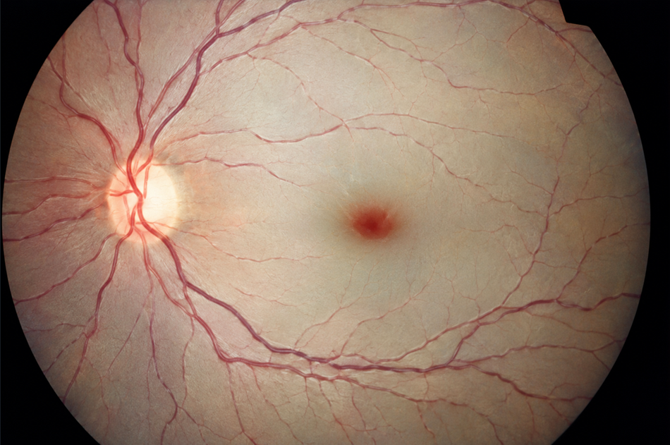
Retinal artery occlusion is one of the most urgent conditions in ophthalmology. It occurs when blood flow to part or all of the retina is interrupted, most often due to an embolism or thrombotic occlusion.
The types are:
- Central retinal artery occlusion – usually causes sudden, painless, and severe vision loss in the affected eye.
- Branch retinal artery occlusion – causes vision loss in a specific part of the visual field, depending on the branch affected.
In central retinal artery occlusion, the entire retina is pale, with a characteristic red spot in the fovea known as a cherry-red spot. It results from the contrast between the ischemic retina and the preserved color of the choroid, which is visible through the thin foveal area.
The condition may be associated with carotid atherosclerosis, cardiac emboli, and other vascular diseases. In patients over 50, giant cell arteritis should also be considered, especially if headache, pain when chewing, or general symptoms are present.
Central retinal artery occlusion is considered a form of acute ischemic stroke of the retina. Immediate referral to an emergency department for evaluation and management under a stroke protocol is necessary. Patients should not wait for a scheduled eye exam or rely on home remedies. Treatment options are limited and depend on the time elapsed and the specific clinical situation.
Hypertensive retinopathy
High blood pressure gradually damages the blood vessels of the retina, progressing through several phases: vasoconstrictive, sclerotic, and exudative. Initially, there are reversible functional changes: narrowing of the retinal arterioles caused by vascular spasm. With more advanced damage, structural changes occur in the vessel walls, and arteries compress veins where they cross. In severe cases, retinal hemorrhages, hard and wet exudates, and swelling of the optic disc occur.
Hypertensive retinopathy has no early symptoms. With advanced changes, vision may become blurred, and if the macula or optic nerve is affected, significant vision impairment may occur.
Treatment focuses primarily on safely and promptly controlling blood pressure under medical supervision. Severe hypertension with signs of acute organ damage requires urgent medical evaluation.
Wet age-related macular degeneration (AMD)
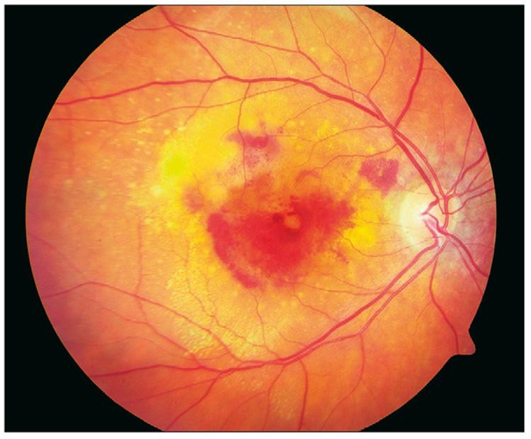
The wet form of AMD is a disease in which abnormal new blood vessels develop under or within the retina, usually originating from the choroid. This condition is called choroidal neovascularization (CNV). The newly formed vessels can leak fluid and blood components, disrupting the structure of the macula and causing blurred vision and distorted images.
Patients may notice that straight lines appear distorted, letters become misshapen, or a dark spot appears in the center of their visual field.
The primary treatment is intravitreal injections of anti-VEGF medications, which block vascular endothelial growth factor and inhibit the formation of abnormal blood vessels. In many patients, treatment can stabilize or improve vision, but repeat injections and regular monitoring are often necessary.
Other retinal vascular diseases
In addition to the conditions listed above, other diseases affect retinal circulation:
- Retinal vasculitis – inflammatory diseases of the retinal blood vessels that may be associated with infections, systemic inflammatory diseases, or other causes.
- Retinal macroaneurysms – localized dilations of retinal arterioles that sometimes cause hemorrhages and swelling.
- Coats disease – a rare condition characterized by abnormally dilated retinal blood vessels and fluid leakage, most often in children and young people, predominantly males.
- Retinopathy of prematurity – associated with abnormal development of retinal blood vessels in premature infants. Severe forms can lead to retinal detachment and vision loss.
- Sickle cell retinopathy – results from vascular abnormalities in sickle cell disease, which can cause peripheral ischemia and abnormal neovascularization.
Diagnosis and treatment of these conditions are determined individually according to the cause, severity, and presence of complications.
What symptoms may indicate retinal vascular disease?
The clinical presentation depends on the type of disease, the area affected, and the extent of vascular damage.
Possible symptoms include:
- sudden or gradual loss of vision;
- blurred central vision;
- distorted images;
- dark spots or missing areas in the field of vision;
- the appearance of floaters, especially with bleeding into the vitreous.
Some diseases, especially early-stage diabetic and hypertensive retinopathy, may cause no noticeable symptoms. Therefore, preventive eye exams are important even when a patient can see well.
When is emergency care needed?
If you experience sudden vision loss or a significant decrease in vision, the sudden appearance of a dark curtain, multiple new floaters or flashes of light, seek immediate medical evaluation. If you suddenly lose vision, do not wait for the symptoms to go away.
It is important to know that these symptoms are not specific to vascular diseases. They may also be signs of other urgent eye conditions, including retinal detachment.
How are retinal vascular diseases diagnosed?
Modern diagnosis combines a clinical eye examination with specialized imaging tests. The choice of tests depends on the suspected disease and the severity of symptoms.
Ophthalmoscopy and fundus photography
After the pupils are dilated, the ophthalmologist examines the back of the eye for hemorrhages, microaneurysms, vascular changes, areas of ischemia, and abnormal new blood vessels. Fundus photography allows findings to be documented and compared at follow-up visits.
Optical coherence tomography (OCT)
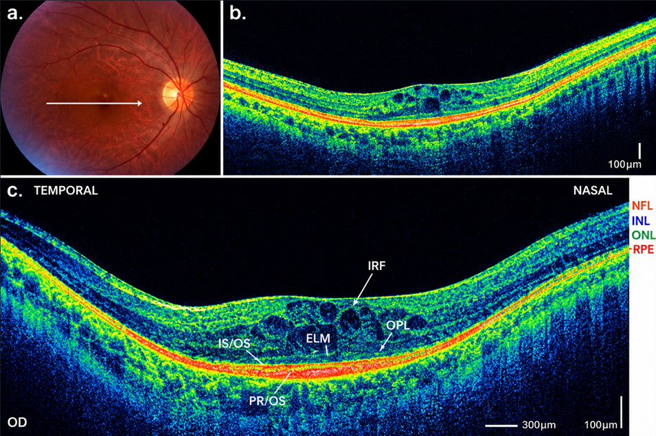
OCT is a noninvasive test that provides detailed cross-sectional images of the retina. It is particularly useful for detecting and monitoring macular edema, intraretinal and subretinal fluid, and structural changes in the macula.
Fluorescein angiography
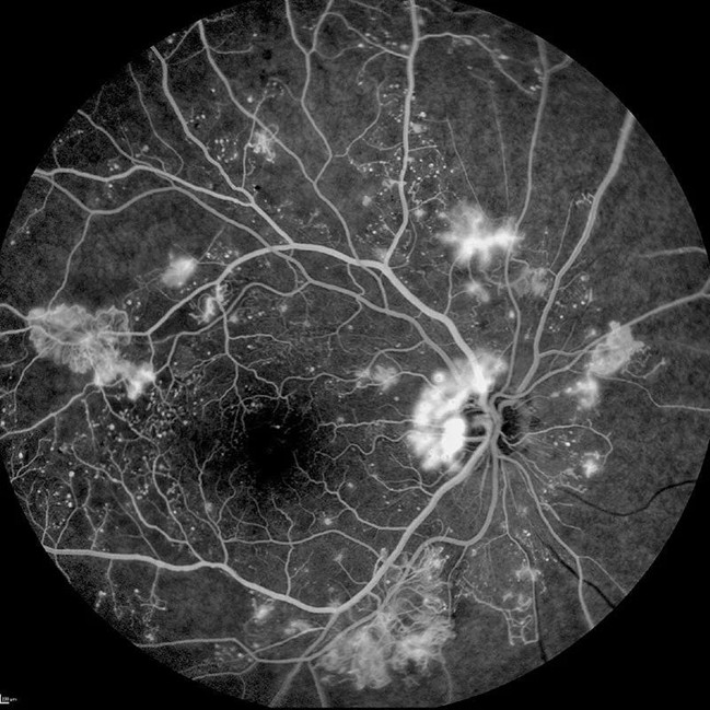
After fluorescein is administered intravenously, the passage of the dye through the retinal vessels is recorded. The method shows vascular leakage, delayed blood flow, areas without blood flow, and neovascularization.
OCT angiography
This method visualizes blood flow in the retinal and choroidal vascular networks without injecting a contrast agent. It is useful for assessing capillary perfusion and abnormal neovascularization, but it does not completely replace fluorescein angiography.
Depending on the clinical situation, additional tests may be needed: blood pressure measurement, blood glucose and glycated hemoglobin tests, a lipid profile, carotid ultrasound, electrocardiography, and a cardiac evaluation.
In cases of acute retinal artery occlusion, the diagnostic evaluation must be arranged urgently and include an assessment for stroke and a cardiovascular source of embolism.
What are the treatment options?
Treatment is tailored to the individual. The goal is to address the cause, limit retinal damage, prevent complications, and improve vision when possible.
Management of systemic diseases
Managing blood glucose, blood pressure, and lipids is an essential part of preventing and treating retinal vascular diseases. In cases of venous occlusion, cardiovascular risk factors should be assessed and any identified conditions treated.
Intravitreal injections of anti-VEGF medications
These medications block the action of vascular endothelial growth factor (VEGF), which stimulates abnormal neovascularization and increased vascular leakage.
Anti-VEGF therapy is a primary treatment for many cases of diabetic macular edema, macular edema following retinal vein occlusion, and wet age-related macular degeneration. The injections are administered into the eye under strict aseptic conditions. Repeated treatments and regular follow-up are often necessary.
Intravitreal corticosteroids
Corticosteroid medications can reduce inflammation and vascular leakage. They are suitable for certain patients with macular edema after the expected benefits and risks have been assessed, including increased intraocular pressure and cataract development.
Laser photocoagulation
Laser treatment is used for proliferative diabetic retinopathy and other cases of retinal ischemia and neovascularization.
Vitrectomy
Vitrectomy is a surgical procedure in which the vitreous is removed. It may be necessary for persistent or recurrent vitreous hemorrhage, tractional retinal detachment, or other severe complications of proliferative diabetic retinopathy.
Treatment for retinal artery occlusion
If central retinal artery occlusion is suspected, immediate referral to an emergency department for evaluation under an acute ischemic stroke protocol is necessary. The appropriate emergency team assesses reperfusion treatment options based on the time since onset, contraindications, and current protocols.
Can retinal vascular diseases be prevented?
Not all retinal vascular diseases can be prevented, but the risk of developing and progressing in many of them can be reduced.
The most important measures are:
- regular monitoring of blood sugar and glycated hemoglobin in people with diabetes;
- keeping blood pressure within recommended limits;
- controlling cholesterol and other lipid levels;
- quitting smoking;
- a balanced diet and regular physical activity;
- routine eye exams, even in the absence of vision problems.
The primary care physician, endocrinologist, cardiologist, and ophthalmologist have complementary roles. Timely communication among them helps improve control of risk factors and enables early detection of complications.
What is the prognosis?
The prognosis depends on the type of disease, the degree of ischemia, whether the macula is affected, the presence of bleeding and neovascularization, and the time until treatment begins.
For some patients, modern treatment can significantly improve vision. For others, the main goal is to preserve remaining visual function and prevent further damage.
Even with successful treatment, permanent visual impairment may remain. In such cases, vision rehabilitation, appropriate optical aids, and psychological support can improve daily functioning and quality of life.
It is important for patients to understand that treatment is often a long-term process. Regular follow-up exams are an essential part of it, even when vision has improved.
New possibilities and future prospects
Ophthalmology is advancing rapidly, and new approaches to more effective and longer-lasting treatment of retinal diseases are being studied.
The main areas of research include:
- Longer-acting anti-VEGF drugs, which may reduce the frequency of injections in suitable patients.
- Gene therapy, being studied as a way to suppress certain disease mechanisms for a longer period.
- Artificial intelligence, which assists in the automated detection of diabetic retinopathy and other retinal changes during screening.
- Improved imaging technologies that allow a more precise assessment of retinal microcirculation and early structural changes.
These approaches have significant potential, but their use depends on the specific disease, scientific evidence, and approved indications. New technologies complement, rather than replace, the ophthalmologist’s clinical judgment.
Questions and Answers
What are the most common retinal vascular diseases?
The main ones include diabetic retinopathy, retinal vein and artery occlusions, and hypertensive retinopathy. Wet age-related macular degeneration is also an important disease associated with abnormal neovascularization.
Are these diseases painful?
Most retinal vascular diseases are painless. Therefore, the absence of pain does not mean there is no serious damage.
Which symptoms require an urgent examination?
Sudden loss or significant reduction of vision, the appearance of a dark curtain, or numerous new floaters require immediate medical evaluation.
Can lost vision be restored?
This depends on the cause and extent of the damage. In some conditions, vision can improve significantly, while in cases of severe ischemia, the damage may be irreversible. Early treatment increases the chances of a favorable outcome.
How often should people with diabetes have eye exams?
At least one dilated eye exam per year is usually necessary. If retinopathy is present, during pregnancy, or under other circumstances that increase risk, the ophthalmologist determines the interval individually.
Are intravitreal injections painful?
The procedure is performed under local anesthesia and usually causes minimal discomfort. Mild irritation or a foreign body sensation may occur temporarily afterward. Severe pain or a sudden worsening of vision after an injection requires immediate contact with the treating doctor.
Can these diseases be prevented?
Not always, but controlling diabetes, blood pressure, and lipid levels, quitting smoking, and having regular eye exams can significantly reduce the risk of damage and progression.
Is retinal artery occlusion related to stroke?
Yes. Central retinal artery occlusion is considered an acute ischemic stroke of the retina and requires immediate evaluation in a stroke emergency department.
Are these diseases hereditary?
Most are not directly inherited, but genetic factors can affect the risk. In some diseases, such as certain forms of macular degeneration, inherited predisposition plays a more significant role.
When should I see a specialist?
If you experience a sudden change in vision, seek care immediately. If you have diabetes, high blood pressure, or other risk factors, have preventive eye exams as recommended by your doctor, even if you have no symptoms.
Conclusion
Retinal vascular diseases are a significant cause of vision problems and can have serious consequences if they go undetected or untreated. Some develop gradually and without symptoms, while others occur suddenly and require emergency medical attention.
Regular preventive eye exams, management of systemic diseases, and timely consultation with an ophthalmologist are essential steps in preserving vision.
Don’t wait for your vision to deteriorate before taking care of it. Early detection can make a significant difference.
Important: This article is for informational purposes and does not replace an eye exam or an individual medical consultation. It is part of the information campaign by Pentagram Eye Hospital, in partnership with the Retina Bulgaria Association, aimed at raising awareness of retinal diseases: “See What Cannot Be Seen.”

Nine Fascinating Facts About Eye Color
Eyes are among a person’s most distinctive features, and their color holds many more fascinating stories than we might imagine..
Read More
Modern Diagnosis of Retinal Diseases
Modern tests for retinal diseases. Color fundus photography, fluorescein angiography, autofluorescence imaging, and optical coherence tomography—when they are used and.
Read More