

What Is Cataract Surgery?


Author: Dr. Milen Lazov, ophthalmologist at Pentagram Eye Hospital
Cataracts are among the most common eye conditions worldwide and a leading cause of vision loss in older adults. The good news is that they can now be treated successfully, and Bulgaria has technologies and specialists on par with European standards. Dr. Veselin Daskalov shared this a week ago on BNR’s podcast “At the Center of the System”. A week ago, Dr. Risto Traykovski also explained why cataract surgery should not be postponed and what complications a delay can cause.
Today, we explore the subject in greater depth with one of our experienced ophthalmologists, Dr. Milen Lazov, who explains the different approaches to cataract surgery, the types of lenses, and what determines the choice of the most suitable lens to replace the eye’s damaged natural lens.
WHAT IS CATARACT SURGERY?
There are three types of cataract surgery:
–Intracapsular extraction /ICCE/ – the oldest and most invasive method; the entire lens is removed together with the capsular bag, and a large corneal incision is made and closed with sutures; it is used when the lens apparatus is severely damaged; the implant is attached to the iris or sclera;
–Extracapsular extraction /ECCE/ – in this method, the capsular bag is preserved and the lens is mechanically removed through a smaller incision, which is closed with sutures; it is also used when the lens apparatus is damaged, the cataract is hypermature, or there is corneal pathology;
–Phacoemulsification – the gold standard worldwide and the method most commonly used at our hospital. It is minimally invasive and allows rapid visual recovery. It is performed under local anesthesia (eye drops), without sutures, through an incision of about 2 millimeters. An opening is made in the anterior capsule (capsulorhexis), and fluid is introduced to separate the capsule from the lens. The lens is then broken up with ultrasound using a phaco machine and suctioned out (phacoemulsification), the lens implant is positioned in the capsular bag, and the incision is closed.
As variations, parts of the surgery can be performed with the help of lasers. Examples include Capsulaser, which performs the capsulorhexis instead of the surgeon, and the Black Victus femtosecond laser, which performs both the capsulorhexis and the initial fragmentation of the cataract, reducing the need for ultrasound and its negative effects. After surgery, an eye patch is applied and removed the next day.
WHAT DETERMINES THE EXPECTED OUTCOME OF SURGERY
First and foremost is the overall condition of the eye. The eye has both an optical part (the cornea, lens, and, to some extent, the vitreous) and a sensory part (the retina, particularly the macula, and the optic nerve), as well as nerve centers and pathways in the brain. Damage to these structures (e.g., corneal dystrophies and opacities, opacities in the vitreous, forms of macular degeneration and macular edema, diabetic retinopathy, vascular or inflammatory diseases, and glaucoma) affects visual function to varying degrees, depending on the individual case.
Also important is the degree of cataract maturity, including complicated and hypermature cataracts. (More on this topic HERE)
In the absence of significant accompanying eye conditions, full visual rehabilitation (100%) is usually achieved after a short recovery period, i.e., the patient will achieve 100% vision, the maximum possible for that individual. In other cases, insofar as the vision loss is due to the cataract itself, the improvement is significant: patients with hypermature cataracts who previously had only light perception achieve form vision—they can see images—but their vision is still not 100%. In patients with macular degeneration, central vision may improve only slightly (this also depends on the form and severity of AMD), but overall and peripheral vision will improve significantly.

DOES THE OPERATION REQUIRE A HOSPITAL STAY
Yes, the operation requires a hospital stay, but not overnight—only for a few hours.
In the first stage, an eye examination, individualized intraocular lens calculation, blood tests, and consultations with internal medicine specialists are performed.
Then, immediately before the operation, the patient is admitted to the ward, and the pupil of the eye to be operated on is dilated; an individual anesthesia consultation is also conducted.
The surgical procedure follows, which takes about 15–20 minutes in uncomplicated cases. After the operation, the patient remains under observation for a short time and then leaves the hospital on the same day in a completely normal condition. It is advisable to have someone accompany you, as driving is absolutely contraindicated after the procedure. However, there are no contraindications to traveling, including to another town or city.
WHAT ARE THE TYPES OF INTRAOCULAR LENSES
In recent years, the intraocular lens has evolved from a means of visual rehabilitation into a method for overcoming the physiological limitations of the eyes.
From the perspective of the patient and their visual abilities after surgery, we can divide them into:
- Providing focus at distance or near: these are monofocal lenses and their enhanced versions (mono +), which have a slightly improved ability to focus at intermediate distances. These lenses provide the clearest vision without side effects at the corresponding distance (near or far), while other visual needs are corrected with prescription glasses.
- Providing focus at two distances (distance and intermediate or distance and near).
This group mainly includes extended depth of focus (EDOF) lenses, bifocal lenses, and some other diffractive lenses. Minimal side effects may be expected, and the third focal range is corrected with glasses. - Providing focus at all distances: these are conventional trifocal lenses and some lenses with an enhanced depth of focus achieved through different technologies. Statistically, the most side effects may be expected with these lenses (e.g., halos around light sources, glare, and starbursts), although these occur relatively rarely or fade over time and are not significant. They depend on the specific optical technology of the lens. It is important that the choice of this type of lens be strictly individualized according to the patient’s lifestyle, expectations, and the characteristics of their eye anatomy and physiology.
- Providing correction of corneal astigmatism – these are toric lenses, which can be versions of any of the lenses listed above. They are positioned at a specific angle in the capsular bag and are used to correct astigmatism.
- –Special types of implants – iris-lenses, pinhole lenses for correcting irregular astigmatism; scleral- or iris-fixated lenses for aphakia. They are used in more complex surgeries involving trauma or complex eye reconstruction, after corneal transplantation, and in patients with keratoconus.
Depending on their structure, lenses can be:
– Rigid or soft (foldable). Almost all lenses are foldable; rigid lenses are used only in very limited cases /ICCE/
– Three-piece or monolithic: depending on the structure of the implant. The former provide better fixation stability and are used for specific indications; the optics are the same in both types.
All modern lenses have an ultraviolet light filter.
– Yellow filter – some lenses have a yellow filter that reflects their ability to block some of the blue light emitted by the screens that are increasingly common in our daily lives. It allows for a smoother transition from the yellow-brown color perception caused by a cataract to clear vision after surgery. However, it also changes fine color perception and contrast.

WHAT DETERMINES THE CHOICE OF AN INTRAOCULAR LENS
Sometimes, the wrong choice of intraocular lens may not only fail to improve visual function but may even worsen it—such as when a lens with complex optics is implanted in an eye with an existing condition.
In other words, lenses with greater capabilities also place greater demands on the capabilities of the eyes in which they are implanted.
- In eyes with a weak lens support system, three-piece lenses are preferred, or a rigid lens may need to be implanted.
- In eyes with an underlying condition (macular degeneration, retinopathy, glaucoma), implantation of a monofocal, clear lens is generally preferred.
At Pentagram Eye Hospital, cataract surgery is performed with a referral under the National Health Insurance Fund, meaning the surgical procedure is free for the patient. Patients pay only for the lens that will be replaced during surgery, their choice of surgical team, and certain supplies related to the procedure.
You can find more information on the topic at:
Dr. Veselin Daskalov – What is a cataract, what causes it, and what are the treatment options?
Dr. Risto Traykovski – Should you wait for a cataract to “ripen,” and what are the consequences of delaying surgery?
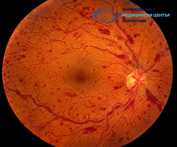
Retinal Vascular Diseases
Retinal vascular diseases. Causes. Symptoms. Diagnosis. Treatment. Emergencies.
Read More
For the first time in Bulgaria and Eastern Europe – intraoperative treatment of glaucoma with the new ELIOS laser
First in Bulgaria: ELIOS laser for the treatment of glaucoma and cataracts at the "Pentagram" clinic. Dr. Daskalov applies a.
Read More