

Ocular Surface Disease – Dry Eye
Author: Dr. Ralitsa Kermedchиеva – ophthalmologist at Pentagram Eye Hospital
Ocular surface disease (OSD) is an umbrella term for conditions affecting the cornea, conjunctiva, and eyelids, with dry eye disease (DED) being the most common form.
Timely diagnosis and treatment of ocular surface diseases is a cornerstone of successful anterior segment surgery and overall eye health.
Understanding and managing these conditions is no longer optional—it is essential for optimal surgical outcomes and patient satisfaction.
Ocular surface disease (OSD) is a complex condition that affects both the tears and the ocular surface, leading to various symptoms such as discomfort, visual disturbances, and tear film instability.
It is characterized by increased tear film osmolarity and inflammation, which can manifest clinically as superficial punctate keratitis, conjunctival hyperemia, and papillary conjunctivitis. (1)
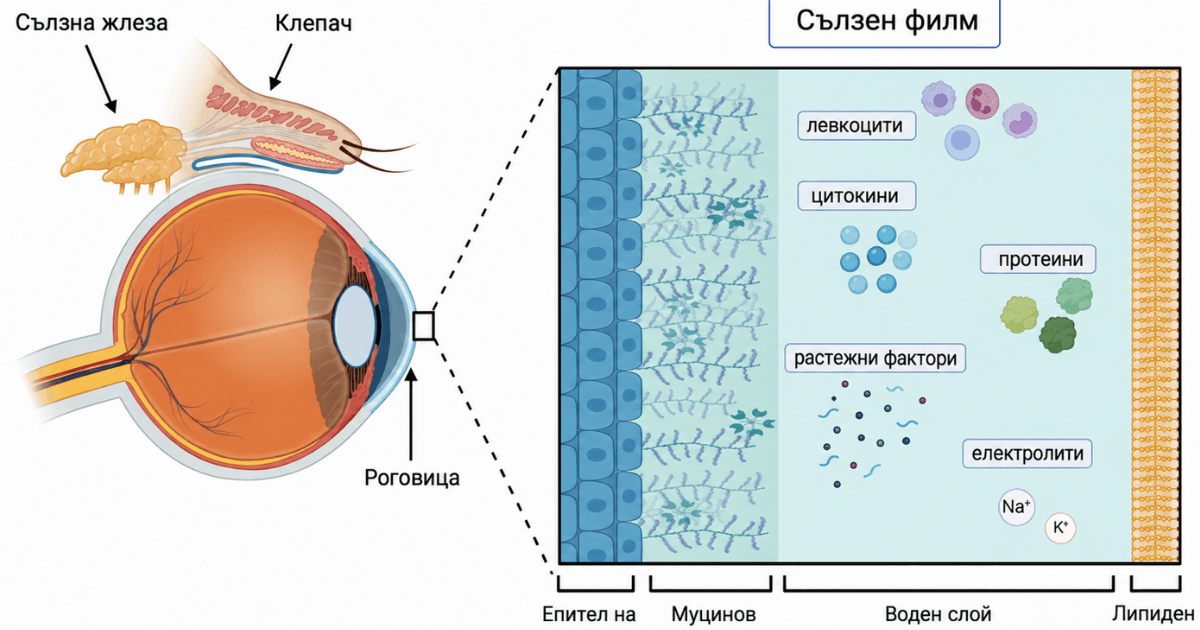
Fig. 1 Anterior ocular surface
Immune-inflammatory mechanisms
- The etiology of OSD is diverse and includes environmental and genetic factors, aging, dry eye syndrome, blepharitis, meibomian gland dysfunction (MGD), and chronic use of eye drops containing preservatives
- Inflammation of the ocular surface is thought to play a key role in the pathogenesis of OSD
- Numerous studies report higher levels of interleukins (IL)-1β, IL-6, IL-8, IL-10, interferon-γ (IFN-γ), and tumor necrosis factor-α (TNF-α) in the tears of patients with DED compared with control groups (2)

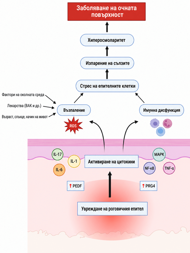
Fig. 2 Immune-inflammatory mechanisms
Dry eye disease (DED)
It is characterized by subnormal tear production, rapid breakdown of the tear film, and accelerated evaporation of tears from the ocular surface
The TFOS DEWS II report suggests that dry eye, regardless of subtype, enters a final common pathway in which hyperosmolarity and inflammation of the ocular surface create a vicious cycle, triggering an inflammatory cascade that leads to reduced expression of glycocalyx mucins and damage to goblet cells. (3)
These changes affect the integrity of the ocular surface and influence all optical measurements
Dry Eye Disease (DED) … Disease, not a syndrome
The Tear Film and Ocular Surface Society (TFOS) Dry Eye Workshop (DEWS) III diagnostic methodology report defines DED as:
“A multifactorial, symptomatic disease characterized by a loss of homeostasis of the tear film and/or ocular surface, in which tear film instability and hyperosmolarity, inflammation, ocular surface damage, and neurosensory abnormalities are etiological factors.” (4)

Fig. 3 Risk factors
Classification
– Predominantly evaporative (impaired lipid layer due to meibomian gland dysfunction (MGD) – 80%
– Aqueous deficient (with reduced tear film volume) – 10%
Meibomian gland dysfunction (MGD) is considered the primary etiological factor in DED.
Definition
MGD is a diffuse, chronic abnormality of the meibomian glands characterized by obstruction of the terminal duct and/or qualitative or quantitative changes in glandular secretion.
This may lead to changes in the tear film, symptoms of eye irritation, clinically visible inflammation, and disease of the anterior ocular surface.

Fig. 4 Pathophysiological mechanisms of MGD
MGD – clinical findings (5)
– Qualitative or quantitative changes in secretion
– Duct obstruction with epithelial hyperkeratinization
– Accumulation of meibum with increased viscosity, leading to increased bacterial growth and release of toxic mediators
– Gland dilation followed by atrophy
– Altered eyelid morphology – thickening and irregularity of the lid margin with hypokeratinization and increased vascularization
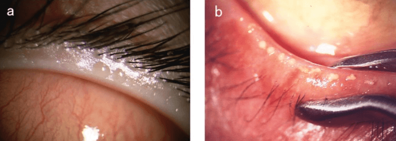
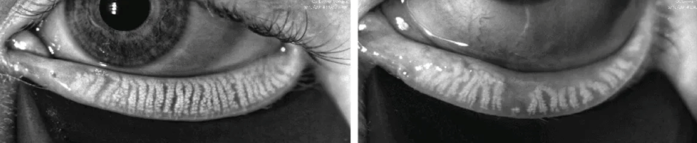
Fig. 5 Meibography – meibomian gland dropout indicates a complete or partial loss of acinar glandular tissue
Cataract surgery and DED
This is the most commonly performed surgical procedure and is also developing as a successful refractive procedure, with patients’ expectations for postoperative outcomes becoming increasingly high.
Inaccurate intraocular lens (IOL) power calculations resulting from changes in the ocular surface can prevent optimal visual outcomes, while cataract surgery can worsen existing DED. (6,7)
According to the literature, 50% of patients undergoing cataract surgery who had meibomian gland dysfunction (MDG) were asymptomatic.
Diagnosing and promptly treating dry eye disease not only improves patient comfort but also enhances postoperative refractive outcomes and quality of vision. (8)
The impact of DED on cataract surgery
Biometry
Accurate biometry requires a stable tear film, the first refractive medium on the ocular surface.
Several studies demonstrate the importance of preoperative ocular surface preparation: (9,10,11)
– Patients with tear film hyperosmolarity have poor repeatability of keratometry values used in IOL calculations compared with patients with normal osmolarity
– The repeatability of both keratometry and axial length (AL) measurements is negatively correlated with TBUT
– In patients with DED, differences in calculated IOL powers can be significant due to tear film instability and a lack of repeatability between measurements: over 0,5D, with a maximum difference of 5,5D
– A study by Roggla et al. focused on the effect of artificial tears on keratometry measurements before cataract surgery, concluding that biometry should be performed 5 minutes after instillation of artificial tears in patients with DED
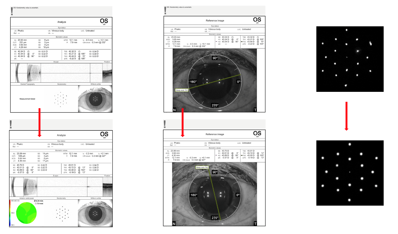
Fig. 6 Biometry – IOL Master – differences in biometric measurements before and after application of an artificial tear (5 minutes)
Diagnosis
– Questionnaire – OSDI
– Schirmer test
– TBUT
– Ocular surface examination – corneal and conjunctival staining
– Osmolarity
– Meibography
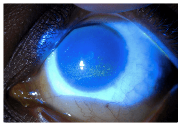
Fig. 7 Fluorescein staining
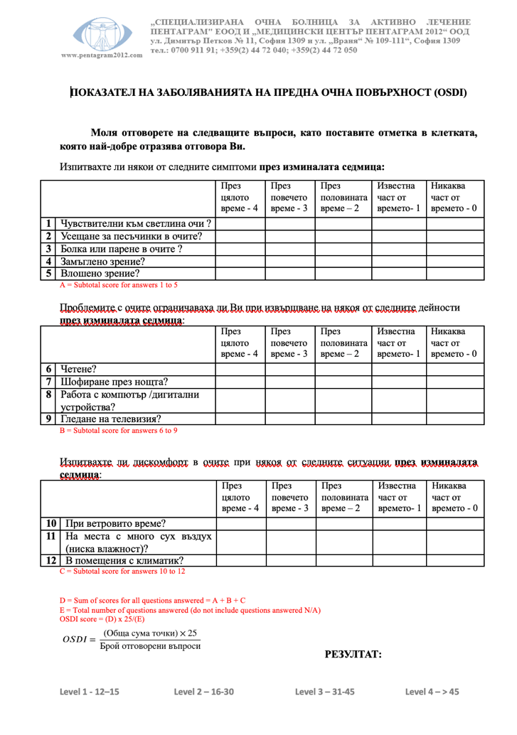
Fig. 8 OSDI questionnaire
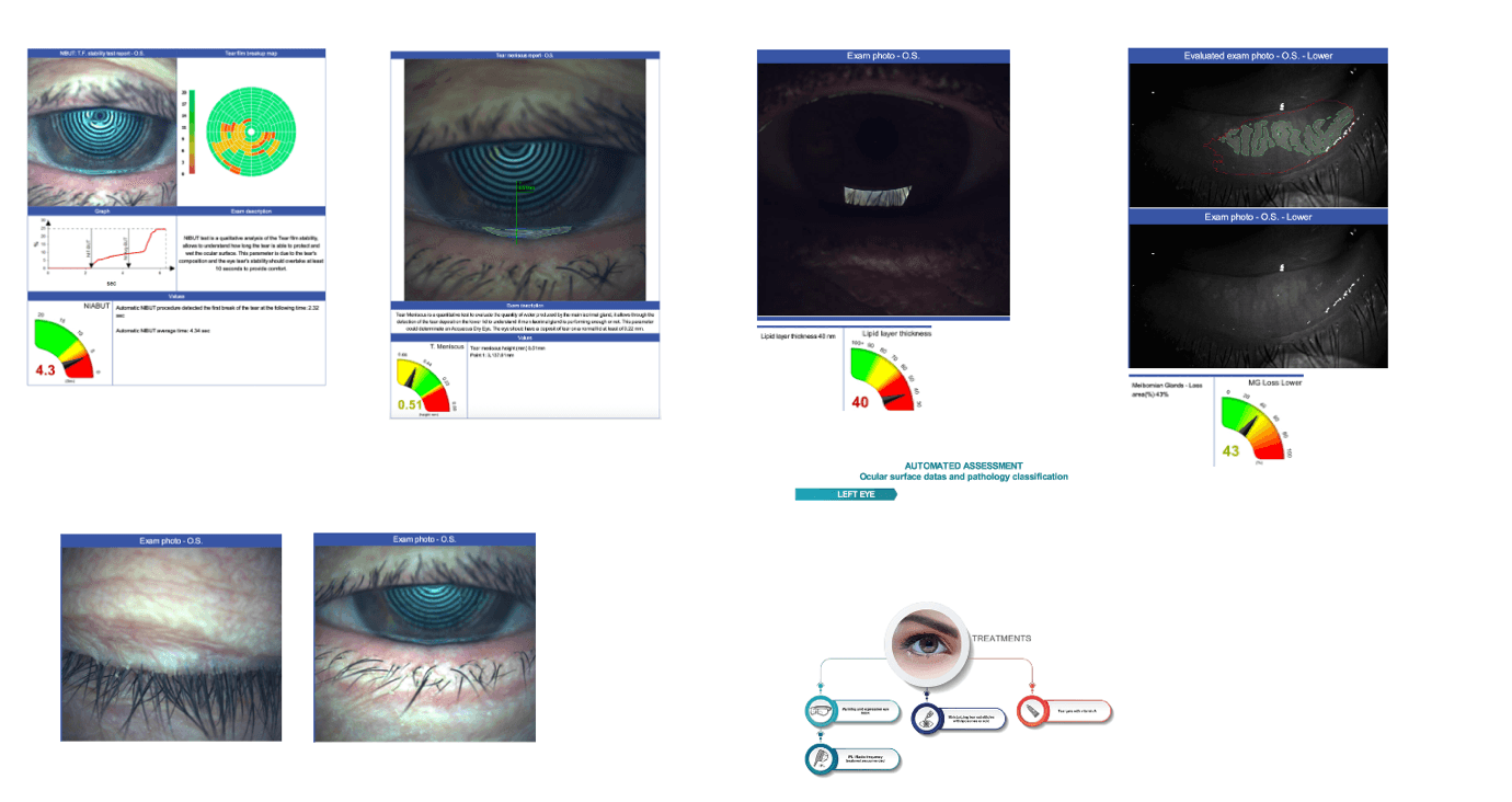
Fig. 9 Tear film assessment and meibography
Optimizing the ocular surface before surgery in patients with DED (12)
– Avoiding epitheliotoxic medications – NSAIDs
– Avoiding eye drops containing benzalkonium chloride (BAK/BKC)
– Treatment with artificial tears and gels
– IPL – treatment of preexisting MGD
– Cyclosporines (Cyclosporine 0.1% Ikervis)
In a study, Hovesian et al. evaluated the effect of preoperatively administered 0,09% cyclosporine on ocular surface regeneration and reported significantly improved accuracy of the postoperative spherical equivalent outcome. Pretreatment of DED also significantly improved clinical signs after surgery. (13)
These results reinforce the fact that adequate treatment of DED plays an important role in performing ocular biometry to achieve optimal visual outcomes.
Risk factors for DED after cataract surgery
Systemic/lifestyle factors
– Age > 60 years
– Female sex
– Diabetes
– Hypertension
– Systemic use of antidepressants, antihistamines
– Infrequent blinking (computer vision syndrome)
Preexisting eye conditions
– DED – worsening, MGD – worsening
– Blepharitis, conjunctivitis, pterygium
– Allergy – increased inflammatory response
– Previous eye surgery – impaired corneal innervation
Mechanisms of postoperative DED
– Surgical trauma – disruption of corneal nerve fibers
– Light exposure from the operating microscope
– Repeated drying and irrigation
– Postoperative therapy – preservatives (damage epithelial cells and goblet cells), NSAIDs (damage goblet cells and affect corneal innervation)
– Postoperative inflammation – increased levels of multiple inflammatory factors (prostaglandins, IL-1, 6, 8, interferon)
In a study following patients after cataract surgery, Giannaccare et al. (14) reported the following results:
– Homeostasis indicators, such as corneal sensitivity, tear film stability, and mean goblet cell density, decreased on the first day after surgery, reaching a peak on day 7, followed by progressive recovery
– Dry eye symptoms persisted for 1-3 months after surgery
MGD is an important but often underestimated factor contributing to the vicious cycle of dry eye, which can worsen postoperatively.
There are several clinical interventions for MGD – IPL with meibomian gland expression, eyelid hygiene, omega-3 fatty acid supplements, and oral antibiotics.
The use of artificial tears plays an important role in controlling inflammation in postoperative DED by improving tear fluid clearance and reducing the concentration of proinflammatory cytokines. Their use should be recommended for all patients for at least the first month after phacoemulsification, as it improves tear film stability and postoperative DED symptoms.
Severe symptoms impair patients’ quality of life and can also compromise visual outcomes after cataract, refractive, and corneal surgery. Timely diagnosis and treatment of the condition will ensure good postoperative visual outcomes and patient comfort.
Reference:
1. Scarpellini et al., The potential role of regulated cell death in dry eye diseases and ocular surface dysfunction. Int. J. Mol. Sci. 2023
2. Roda, M.; Corazza, I.; Bacchi Reggiani, M.L.; Pellegrini, M.; Taroni, L.; Giannaccare, G.; Versura, P. Dry eye disease and tear cytokine levels-a meta-analysis. Int. J. Mol. Sci. 2020,
3. Willcox M, Argueso P, Georgiev G, et al. TFOS DEWS II tear film report. Ocul Surf. 2017
4. Wolffsohn JS, Benítez-Del-Castillo JM, Loya-Garcia D, Inomata T, Iyer G, Liang L, Pult H, Sabater AL, Starr CE, Vehof J, Wang MTM, Chen W, Craig JP, Dogru M, Perez VL, Stapleton F, Sullivan DA, Jones L; + TFOS collaborator group. TFOS DEWS III: Diagnostic Methodology. Am J Ophthalmol. 2025 Nov;279:387-450. doi: 10.1016/j.ajo.2025.05.033. Epub 2025 May 30. PMID: 40451408.
5. Tomlinson A, Bron AJ, Korb DR, et al. The international workshop on meibomian gland dysfunction: report of the diagnosis subcommittee. Invest Ophthalmol Vis Sci. 2011;52(4):2006–2049. doi: 10.1167/iovs.10-6997f
6. Gupta et al., J Cataract Refract Surg. 2018
7. Trattler et al., Clin Ophthalmol. 2017
8. Cochener et al., J Cataract Refract Surg. 2018
9. Röggla V, Leydolt C, Schartmüller D, et al. Influence of artificial tears on keratometric measurements in cataract patients. Am J Ophthalmol. 2021;221:1–8. doi:10.1016/j.ajo.2020.08.024
10. Epitropoulos AT, Matossian C, Berdy GJ, Malhotra RP, Potvin R. Effect of tear osmolarity on repeatability of keratometry for cataract surgery planning. J Cataract Refract Surg. 2015;41(8):1672–1677. d
11. Hiraoka T, Asano H, Ogami T, et al. Influence of dry eye disease on the measurement repeatability of corneal curvature radius and axial length in patients with cataract. J Clin Med. 2022;11(3):710. doi
12. Labetoulle M, Rousseau A, Baudouin C. Management of dry eye disease to optimize cataract surgery outcomes: two tables for a daily clinical practice. J Fr Ophtalmol. 2019;42(8):907–912. doi:10.1016/j.jfo.2019.03.032.
13. Hovanesian J, Berdy G, Epitropoulos A, Holladay J. Effect of cyclosporine 0.09% treatment on accuracy of preoperative biometry and higher order aberrations in dry eye patients undergoingcataract surgery. Clin Ophthalmol. 2021;15:3679–3686. doi:10. 2147/OPTH.S325659.
14. Giannaccare, G.; Barabino, S.; Di Zazzo, A.; Villani, E. Preventing and Managing Iatrogenic Dry Eye Disease during the Entire Surgical Pathway: A Study Focusing on Patients Undergoing Cataract Surgery. J. Clin. Med. 2024, 13, 748.

PRESBYOPIA vs HYPEROPIA – what is the difference?
Farsightedness What is important to know? Farsightedness (hypermetropia) is a common congenital vision condition. It represents a refractive error of.
Read More
Occlusion of the Central Retinal Artery (CRAO) is the Eye’s Analog of Ischemic Stroke
Occlusion of the Central Retinal Artery (CRAO): symptoms, risk factors, diagnosis, and emergency treatment for sudden painless vision loss.
Read More