

Age-related macular degeneration causes irreversible vision loss in older adults

Author: Dr. Teodor Daskalov
Age-related macular degeneration (AMD) is one of the most common diseases that cause significant and irreversible loss of central vision in older adults. Early detection and modern treatments can significantly slow the progression of the disease.
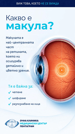
The macula is the central part of the retina that allows us to see fine details, read, recognize faces, and perform activities requiring precise central vision. The disease usually develops after age 50, and its prevalence increases significantly with age. In the early and intermediate stages, patients often have no noticeable symptoms or experience gradual changes in vision. This is why regular preventive eye exams are especially important.
How often does AMD progress?
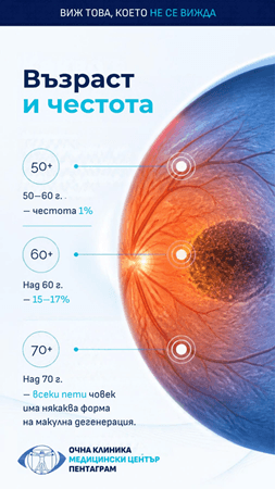
In many patients, the disease remains in an early or intermediate stage for years. However, the risk of progression to advanced AMD depends on the changes present in the macula and the condition of the other eye. Over 5 years, the risk can range from approximately 1.3% in patients with small drusen and minimal pigment changes to about 43% in those with advanced AMD in the other eye or significantly reduced visual acuity due to AMD. Large drusen, geographic atrophy, and existing advanced AMD in one eye are among the most important clinical signs of a higher risk of progression. The prevalence of advanced forms also increases with age—approximately 1% among people aged 65–74 and about 5% among those aged 75–84.
What are the risk factors?
Age is the most important risk factor, but the development and progression of AMD result from the interaction of multiple factors. Well-established risk factors include:
- family history and genetic predisposition;
- certain genetic variants, including in CFH and ARMS2/HTRA1;
- smoking;
- age.
For some factors, including high blood pressure, elevated BMI, and iris color, the evidence is less clear.
Dry and wet forms of macular degeneration
AMD is traditionally classified as neovascular (“wet”) or non-neovascular (“dry”), with the dry form being considerably more common.
Dry AMD
In the dry form, structural changes gradually develop in the retina–retinal pigment epithelium–Bruch’s membrane complex, often with drusen and pigment abnormalities. In some patients, the condition may progress to geographic atrophy, in which retinal cells and photoreceptors in the macular area are lost.
Dry AMD usually progresses more slowly than wet AMD, but it can lead to significant and irreversible loss of central vision. In certain patients with intermediate or advanced dry AMD, specially selected nutritional supplements based on the AREDS2 (Age-Related Eye Disease Studies) formula can reduce the risk of progression to an advanced stage. They do not restore vision that has already been lost or prevent the disease in all patients, so their use should be based on the stage of AMD and individual risk.
Drug therapies are now available to slow the progression of geographic atrophy, which changes the traditional understanding that dry AMD is entirely untreatable.
In recent years, a new approach to treating dry AMD has emerged – photobiomodulation (PBM).
One of the most interesting technologies in this field is the Valeda Light Delivery System, which uses a combination of light at wavelengths of 590, 660, and 850 nm. The aim is to influence cellular metabolic processes in the retina and support retinal cell function through controlled light stimulation. Unlike intravitreal therapy for wet AMD, photobiomodulation is a noninvasive treatment and does not require injection of medication into the eye. It is a promising additional approach for certain patients, but it does not yet replace established standards for monitoring and treatment.
What do clinical studies show?
The results of LIGHTSITE III, a randomized controlled study in patients with intermediate dry AMD, show improvements in visual function and certain anatomical measures in patients treated with photobiomodulation.
The more recent 24-month analysis of LIGHTSITE III, published in 2026, continues to investigate the long-term efficacy and safety of the therapy. In the study, patients received a series of 9 treatments, three times a week over a period of 3–5 weeks, with the treatment course repeated every 4 months. These results are promising, but photobiomodulation should be presented as an innovative treatment option for dry AMD, not as a substitute for proven approaches or as a treatment that guarantees vision recovery.
Wet AMD
In the wet form, neovascularization develops: abnormal new blood vessels appear under or within the retina. They are fragile and leaky and can cause fluid buildup, bleeding, and subsequent damage to the photoreceptors.
Without timely treatment, the process can lead to rapid and permanent loss of central vision and the formation of fibrous tissue and scars in the macula.
What are the symptoms?
Early changes may go unnoticed. As the disease progresses, the following symptoms may appear:
– gradual or sudden loss of central vision;
– difficulty reading;
– need for more light;
– difficulty recognizing faces;
– distortion of straight lines, known as metamorphopsia;
– appearance of a central spot or a missing part of the image;
– changes in color perception;
– difficulty adapting to darkness.
It is especially important to note that wet AMD can begin with minimal noticeable symptoms, while structural changes are already visible during a retinal examination.
How is it diagnosed?
Diagnosis begins with a comprehensive eye examination and an examination of the back of the eye after the pupils are dilated. Modern diagnosis, however, relies heavily on high-resolution optical coherence tomography (OCT). It allows visualization of even minimal amounts of intraretinal or subretinal fluid, changes in the retinal layers and pigment epithelium, and signs of neovascular activity.
When necessary, additional tests may be used, including OCT angiography (OCTA) and fluorescein angiography, especially when more precise characterization of the abnormal vessels is needed.
How is wet AMD treated?
The major change in the treatment of wet AMD in recent years has been the introduction of anti-VEGF therapy. Anti-VEGF medications inhibit the action of vascular endothelial growth factor (VEGF), thereby reducing abnormal neovascularization, fluid leakage, and macular edema.
Treatment is administered through intravitreal injections into the eye under sterile conditions and local anesthesia. The procedure is performed on an outpatient basis, and the patient can usually go home immediately afterward. It is important to know that treatment for wet AMD is not a one-time procedure. Many patients need repeated injections and long-term follow-up.
The intervals between treatments are determined individually based on disease activity and OCT findings. Anti-VEGF therapies used include aflibercept, faricimab, and brolucizumab, as well as other approved treatment options depending on the specific clinical situation.
Why is early detection so important?
With AMD, timing is especially important. Changes in the early and intermediate stages may be minimal, and patients may not notice a significant decline in vision. However, once abnormal blood vessels develop, the disease can progress much more rapidly.
Regular preventive eye exams, especially after age 50–60 and when risk factors are present, allow AMD to be detected before severe and irreversible vision loss occurs. In patients already diagnosed with AMD, monitoring both eyes is especially important, as the risk of progression is not the same for all patients.
When is it an emergency?
Wet AMD can develop quickly, and damage to central vision is often irreversible. Therefore, certain changes should not be ignored.
See an eye doctor the same day or the next day if you notice:
– sudden distortion of straight lines, such as door frames, lines of text, or window edges;
– a new dark, blurry, or missing spot in the center of your visual field;
– a sudden worsening of central vision in one eye;
– sudden difficulty reading or recognizing faces.
Do not wait for the symptoms to go away on their own. If you cannot quickly schedule an appointment with a specialist, go to an emergency eye clinic. The sooner treatment for wet AMD begins, the greater the chance of preserving vision.
Seek help immediately if you also experience:
– sudden flashes of light in your vision;
– a sudden increase in floaters;
– a “shadow” or “curtain” covering part of your visual field.
These symptoms are not typical of AMD, but they may indicate retinal detachment, which also requires immediate care.
Check your vision at home
Amsler grid
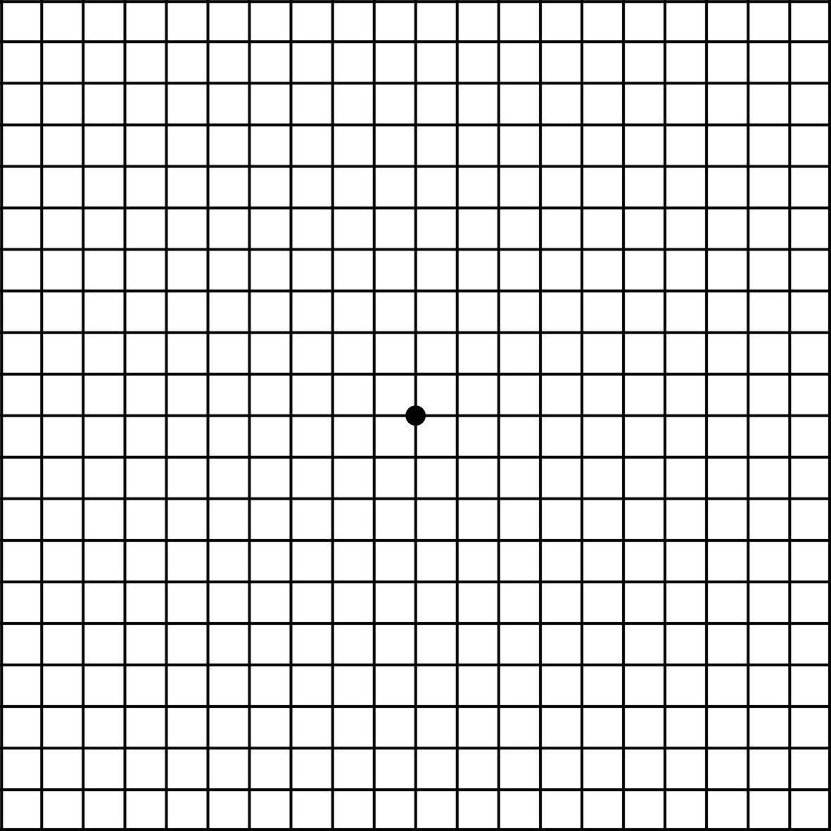
If you have AMD or risk factors, check each eye separately: cover the other eye and look at an object with straight lines or at an Amsler grid. Do this regularly, for example, once a week. The other eye often compensates, so a change may go unnoticed if you look with both eyes.
Important: A home check does not replace a routine eye exam. The absence of symptoms does not rule out disease, because early changes in the macula can occur without symptoms.
How to check your vision with an Amsler grid
An Amsler grid is a square of straight lines with a dot in the center. It helps detect early changes in central vision, such as distortion or missing areas. The check takes about a minute.
How to do it
- Place the grid at eye level, about 30-35 cm from your face, in a well-lit area.
- If you wear reading glasses or bifocals, put them on.
- Cover one eye with your palm, without applying pressure.
- With the other eye, look steadily at the dot in the center of the grid without shifting your gaze.
- Notice whether all the lines look straight and even, whether all the squares are visible and the same size, and whether there are any blurry, dark, or blank areas.
- Repeat with the other eye.
When to be concerned
– some lines look wavy, crooked, or broken;
– part of the grid is blurry, dark, or missing;
– the squares in one area look smaller, larger, or distorted.
What to do if you notice a change
Do not try to attribute it to fatigue or lack of sleep, and do not wait for it to go away. Make an appointment with an eye doctor that same day or the next day and say that you have new distortion or a spot in your central vision. If you notice the change after you have already been diagnosed with AMD, report it immediately, even if it is not yet time for a scheduled exam.
How often
If you have risk factors or have been diagnosed with AMD, check each eye separately once a week. Your doctor may recommend checking more often. Do it at the same time and in the same place so you can compare the results.
Important: The Amsler grid is a self-monitoring tool. It does not provide a diagnosis or replace an eye exam, and a normal result does not rule out AMD. Early changes often cause no symptoms and are detected only during an exam and OCT imaging.
Why Pentagram Eye Hospital?
At Pentagram Eye Hospital, we have many years of experience in diagnosing and treating macular diseases and have been providing intravitreal injection therapy since 2012.
Our approach focuses not only on treating damage that has already occurred, but also on early detection and individualized monitoring for each patient.
We have advanced retinal imaging, including high-resolution OCT technology, which allows precise monitoring of structural changes and assessment of disease activity. This is especially important when determining whether and when further treatment is needed.
The clinic provides current drug therapies for wet AMD. The treatment plan is tailored to each patient based on the type and activity of the disease, the condition of the other eye, and the response to previous treatment. Pentagram also has experience conducting clinical trials of new therapies for retinal diseases, allowing us to be part of the development of modern treatments for macular degeneration.
Important: This article is for informational purposes and does not replace an eye exam or an individual medical consultation. It is part of an awareness campaign by Pentagram Eye Hospital, in partnership with the Retina Bulgaria Association, aimed at raising awareness of retinal diseases – See What Can’t Be Seen.

Screens Increase the Risk of Nearsightedness in Children
Regular preventive eye exams are especially important for children at the beginning of the school year, as vision directly affects.
Read More
Four Ophthalmologists from Pentagram Eye Hospital Are Now in the League of “24 Chasa” Newspaper
There are now four ophthalmologists from the “Pentagram” Eye Hospital in the “24 Hours” newspaper league – “Doctors We Trust”..
Read More